Oosight® Imaging System
Adding Oosight™ to your lab can improve success by giving you a quantitative and reproducible method to measure biological disruption in either fresh or previously frozen oocytes. You can now select oocytes for ICSI and embryos for implantation, and use the system to help improve enucleation efficiency.
Understanding the oocyte is critical to understanding embryogenesis, and studies show that a disrupted spindle apparatus or a weakened zona pellucida in the oocyte can yield lower pregnancy rates. In fact, it has been shown that pregnancy is up to 8 times more likely when the inner zona pellucida is well-ordered.
Our unique and patented solid-state, liquid crystal technology is an easy add-on to your ICSI workstation. Oosight software runs on your computer to capture, display, and analyze your images. Snap an image and click a button to report the data. Meaningful data on molecular order within the sample are organized into an intuitive, exportable report. It’s really that simple.
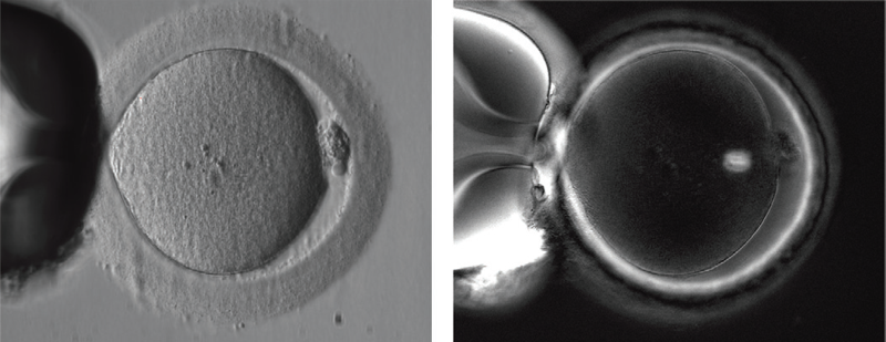
In a conventional contrast image (left) of a human MII oocyte taken just prior to ICSI, structures such as the spindle and multiple layers of the zona pellucida remain invisible. In an Oosight image (right) the spindle is clearly seen to be nicely barrel shaped and the three layers of the zona pellucida are all visible.
Product facts and notices
Key Benefits
- Unprecedented Resolution
High-contrast live images of the oocyte and spindle - Non-invasive Imaging
Does not require the use of any labels or stains, preserving the biology of the spindle and related structures - Quantitative Analysis
Tracks oocyte behaviour over time and automatically records data points of molecular density and orientation - Proven
Successfully used to image many different mammalian species for both enucleation and developmental studies
Features
- Quantitative measurements of molecular order and alignment of birefringent structures, including the spindle, zona pellucida, oolemma, sperm head and tail.
- Calibrated and reproducible data.
- New automated Spindle- and Zona-Finder™ tools.
- New laser targeting tool.
- Reporting capability.
- Seamless integration with most third-party laser systems.
- Foot-pedal controls for quick and easy imaging.
- Models to fit most inverted microscopes.
- High-resolution CCD camera for both retardance-based and general-purpose imaging.
- New smaller footprint for better integration into small work spaces.
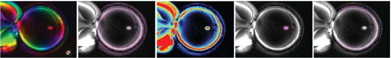
Oosight software visualization tools include (left to right): slow-axis orientation color map, orientation vector overlay, retardance color map, automated SpindleFinder™, and automated ZonaFinder™.
Oosight Imaging System is for research purposes only.
Oosight™ can be used for many applications within the transgenics, stem cell, developmental biology and animal ART sectors, including:
- Spindle imaging for enucleation during nuclear transfer (SCNT)
- Identifying spindle orientation during ICSI
- Assessing egg health and maturation for ART procedures
- Contributing useful data for embryo grading
- Detecting weak points of molecular order in the zona, either for assisted hatching and/or biopsies
- Quantitative imaging of oocytes and embryos for developmental studies
Screening with Oosight Can Make All the Diference
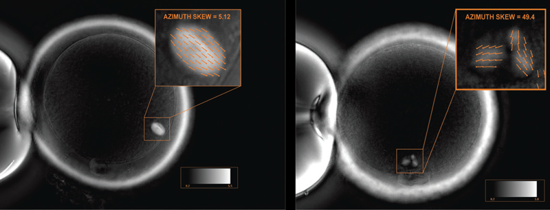
Oosight enables you to determine which subpopulation of oocytes are at high risk for producing chromosomally abnormal embryos. Approximately 1 in 20 cycles contains oocytes that are immature but are nevertheless falsely labeled MII using conventional imaging techniques. Oosight can prevent the potentially damaging effects that result from injecting immature oocytes. The system can also help screen for oocytes with highly disrupted spindles, such as those that are multi-polar.
Improve the efficiency of Cryopreservation
Whenever an application is known to alter the state of the biological material being used, it is imperative that checks and balances are in place to monitor the extent of that change. Oosight can help do this for cryopreserved oocytes by providing a method that helps ensure that vital structures in the oocytes are re-formed to their original pre-frozen state.